Case 01
Diabetic Foot Ulcers: Treatment Journey
A step-by-step visual guide through wound care, healing, and full recovery
PATIENT & CAREGIVER GUIDE
Case overview
Case 01 — overview
- Case Number
- Case 01
- Case Title
- Diabetic Foot Ulcers: Treatment Journey
- Clinical Condition / Diagnosis
- Diabetic foot ulcer
- Patient Demographics
- Not clearly visible in the source slides
- Wound Type
- Deep laceration (~3–4 cm)
- Wound Location
- Foot — as shown in the clinical photographs
- Documented Phases
- 6 phases (Phase 1 to Phase 6)
- Documented Outcome
- Complete Recovery — skin smooth, normal color and texture fully restored
All information on this page is transcribed exactly as presented in the source slides (Avalon Pharma — slide deck: title slide and “From Injury to Full Recovery” slide). Items not shown in the slides are marked “Not clearly visible in the source slides”. No data has been added, estimated or inferred.
Clinical assessment
Wound assessment & findings
- Initial Wound Assessment
- Deep laceration (~3–4 cm). Primary sutures placed to close the wound.
- Wound Dimensions
- ~3–4 cm (laceration length, as stated in the slide)
- Clinical Findings — Phase 2
- Cleaner margins, dark crust forming along suture line.
- Clinical Findings — Phase 3
- Shallow abrasion with yellowish crusts and surrounding erythema.
- Clinical Findings — Phase 4
- Crusts less prominent. Surrounding inflammation subsiding.
- Clinical Findings — Phase 5
- Almost healed. Small faint pink area of new skin visible.
- Clinical Findings — Phase 6
- Skin smooth, normal color and texture fully restored.
Treatment
Intervention documented
- Interventions Documented
- Primary suture closure; wound debridement and re-suturing; suture removal; wound cleaning
- Product / Treatment Used
- Not clearly visible in the source slides
- Application Protocol / Frequency
- Not clearly visible in the source slides
- Dressing Details
- Not clearly visible in the source slides
Healing progression
From Injury to Full Recovery
Six phases of healing — each image shows real progress toward restored skin.
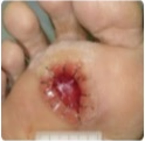
Initial Injury
Deep laceration (~3–4 cm). Primary sutures placed to close the wound.
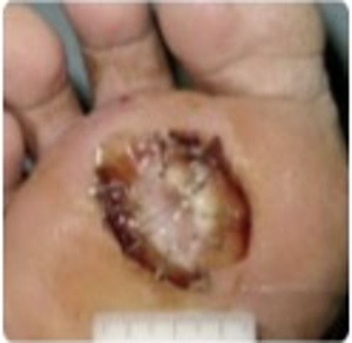
Debridement
Wound debrided and re-sutured. Cleaner margins, dark crust forming along suture line.
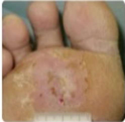
Suture Removal
Sutures removed. Shallow abrasion with yellowish crusts and surrounding erythema.
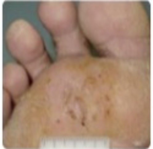
Wound Cleaning
Crusts less prominent. Surrounding inflammation subsiding.
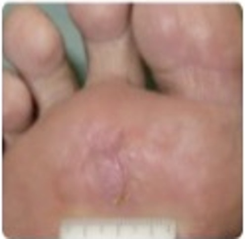
Near Full Healing
Almost healed. Small faint pink area of new skin visible.
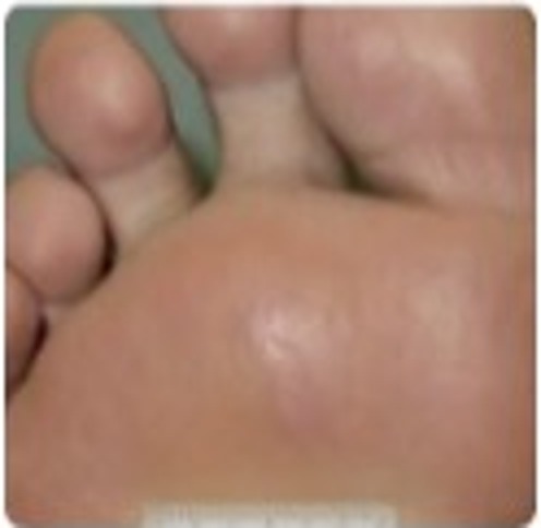
Complete Recovery
Skin smooth, normal color and texture fully restored.
Outcomes
Before & after
Before — Phase 1 (Initial Injury)
Deep laceration (~3–4 cm). Primary sutures placed to close the wound.
After — Phase 6 (Complete Recovery)
Skin smooth, normal color and texture fully restored.
Follow-up
Follow-up record
- Follow-up Schedule / Dates
- Not clearly visible in the source slides
- Follow-up Intervals
- Not clearly visible in the source slides
- Progression Recorded
- Photographic documentation across six sequential phases
- Case Status
- Complete Recovery (Phase 6)
Key findings
Documented key findings
Finding 1
Healing documented across six sequential phases from initial injury to complete recovery.
Finding 2
Wound managed by primary suture closure, followed by debridement and re-suturing.
Finding 3
Sutures removed at Phase 3, with shallow abrasion, yellowish crusts and surrounding erythema recorded.
Finding 4
Surrounding inflammation subsided by Phase 4; crusts less prominent.
Finding 5
Complete recovery documented at Phase 6 — skin smooth, normal color and texture fully restored.
Data completeness
Not shown in the source slides
- —Patient demographics (age, sex, comorbidities)
- —Dates, follow-up intervals and total healing duration
- —Product name, application protocol and frequency
- —Graphs, charts, tables, percentages or statistical data
- —Study, reference or citation information
No graphs, charts, tables, percentages, statistics or study references were present in the supplied slides; none have been recreated. Source: Avalon Pharma — slide deck: title slide and “From Injury to Full Recovery” slide.