Final case template — populated from Case 01
Diabetic Foot Ulcers: Treatment Journey
A step-by-step visual guide through wound care, healing, and full recovery
This template mirrors the documented Case 01 record shown in the Clinical Case Library, the Chronic Wounds category and the case detail page. Open the full case detail.
Fields
Case profile
Documented healing progression
- Phase 1 — Initial Injury: Deep laceration (~3–4 cm). Primary sutures placed to close the wound.
- Phase 2 — Debridement: Wound debrided and re-sutured. Cleaner margins, dark crust forming along suture line.
- Phase 3 — Suture Removal: Sutures removed. Shallow abrasion with yellowish crusts and surrounding erythema.
- Phase 4 — Wound Cleaning: Crusts less prominent. Surrounding inflammation subsiding.
- Phase 5 — Near Full Healing: Almost healed. Small faint pink area of new skin visible.
- Phase 6 — Complete Recovery: Skin smooth, normal color and texture fully restored.
Key findings
- Healing documented across six sequential phases from initial injury to complete recovery.
- Wound managed by primary suture closure, followed by debridement and re-suturing.
- Sutures removed at Phase 3, with shallow abrasion, yellowish crusts and surrounding erythema recorded.
- Surrounding inflammation subsided by Phase 4; crusts less prominent.
- Complete recovery documented at Phase 6 — skin smooth, normal color and texture fully restored.
Photography
Visual timeline
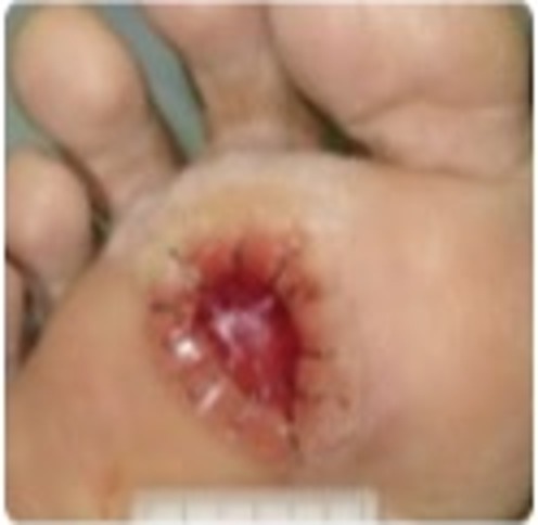
Deep laceration (~3–4 cm). Primary sutures placed to close the wound.
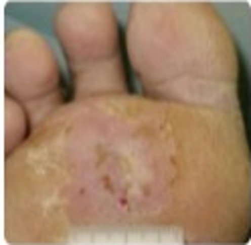
Sutures removed. Shallow abrasion with yellowish crusts and surrounding erythema.
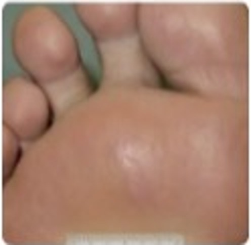
Skin smooth, normal color and texture fully restored.
Source: Avalon Pharma — slide deck: title slide and “From Injury to Full Recovery” slide. Information not shown in the source slides is marked “Not clearly visible in the source slides” and has not been estimated. Medical Affairs | Confidential — For internal use only.